Siibra: a software tool suite for realizing a Multilevel Human Brain Atlas from complex data resources
Nature.com·July 20, 2026
AI Summary
Siibra is a software suite designed to integrate diverse human brain atlases by linking data from multiple imaging modalities and resolution levels. This tool enables the creation of a comprehensive Multilevel Human Brain Atlas that unifies complex neuroimaging resources.
Computational technology opens new possibilities toward understanding the complexity of the human brain, but it requires integrating measurements from different modalities and scales in an anatomical context and exposing them in an interoperable, actionable form. Especially with growing big data resources, accessing information from different scales and modalities coherently for visual exploration, reproducible analysis and application development remains challenging. Here we present siibra, a tool suite that connects diverse data from cloud resources to reference atlases and coordinate spaces. It supports different use cases by making contents accessible through a web viewer, Python library and HTTP application programming interface. Using siibra we implemented a Multilevel Human Brain Atlas linking macro-anatomical concepts and their inter-subject variability with measurements of the microstructural composition and intrinsic variance of brain regions, building on cytoarchitecture as a reference and supporting MRI-based and microscopic templates. The atlas is integrated with the EBRAINS research infrastructure. All software and content are openly accessible.
Decoding the human brain requires consideration of multiple levels of structural and functional organization, using measurements obtained at different spatiotemporal scales with complementary methods. To describe the composition and intrinsic variance of brain regions in conjunction with brain-wide neural network connectivity and inter-subject variability, macro-anatomical concepts need to be linked with microstructural features at a cellular resolution. The integration of such comprehensive information requires reference atlases providing consistent and accurate definitions of anatomical structures across a range of spatial resolutions and modalities in multiple reference coordinate systems, and a consistent model for diverse representations of locations in the brain (for example, brain areas, coordinates, or bounding boxes) that reflect how measurements are linked to anatomy. Such integration of reference atlases with datasets across scales and modalities constitutes a multilevel atlas. Its implementation builds on existing concepts, such as those introduced in ref. 1, and imposes new conceptual and technical difficulties. In particular, it must accommodate rapidly evolving computational technologies and automated workflows to ensure efficient collection and processing of the comprehensive information it offers. Although originating from very different data sources, elements of the multilevel atlas must be exposed in well harmonized, interoperable and actionable formats to make them available in a computationally scalable fashion.
Full story reconstructed from Nature.com. Formatting and media may differ from the original.
Ample data resources exist that describe different aspects of brain organization2, and a broad range of reference atlases are available reflecting complementary principles of brain organization, typically provided in the form of MRI-scale image volumes or labeled surface meshes at spatial resolutions in the millimeter range derived from in vivo imaging. Integration of information into neuroscience workflows has so far been mostly addressed by casting high-resolution measurements into data representations for macroscopic analyses3,4, such as interpolated and averaged feature distributions in surface and voxel formats. Such derivative data make high-resolution features accessible and computable but may reduce the rich regional variance and spatial specificity encoded in the original measurements. In fact, the neuroscience community is increasingly motivated to share primary data with a full level of detail and enable their reuse5. This is supported by the increasing attention of publishers to this topic, leading to a growing abundance of large primary data resources on different online repositories. However, accessing and using data resources from different scales and modalities at their full level of detail remains a technical challenge. An important reason for this is the lack of coherent spatial and semantic integration. The structural relationships between, for example, microscopic measurements from histological studies and whole-brain neuroimaging measurements are often unclear, and the data may refer to different versions and representations of information in reference atlases. In addition, links between measurements and brain areas, such as the label indices in a parcellation map, get easily lost during analysis workflows. Moreover, the consistent interpretation of spatial locations in the brain requires accounting for localization accuracy, different spatial resolutions, and effects of transformations between reference spaces. Another difficulty is the fundamental difference in handling big data, such as whole brain sections at full resolution and three-dimensional (3D) volumes from microscopic studies in the giga- to terabyte range, as compared to data being shared in the form of small files.
Progress in obtaining and analyzing large datasets and the need to link the different modalities across spatial scales imply a paradigm shift from downloading files to interacting with cloud resources and services. Researchers are confronted with new and variable data representations and substantially different tool chains. Therefore, to enhance usability and facilitate data use, we need software solutions that expose data from large cloud resources together with classical file-oriented representations in a unified and interoperable fashion and provide operational principles to connect them with workflows for analysis and modeling.
Here we present siibra (‘software interfaces for interacting with brain atlases’), a tool suite designed to tackle these challenges by offering an informatics framework that integrates data from different modalities and resources with reference atlases spanning macroscopic and microscopic resolutions. The tool suite includes an interactive 3D web viewer (siibra-explorer), an HTTP application programming interface (API; siibra-api) and a Python library (siibra-python). It enables us to specify and process various forms of locations in the brain and assign them to brain areas defined in reference atlases through multilayered semantic and spatial relationships, enabling anatomical characterization and multimodal profiling of regions of interest. Relationships between data, spatial locations and brain areas are modeled as associations with a quantifiable degree of uncertainty and evaluated according to accepted reference coordinate systems and brain region taxonomies. We have developed siibra to implement a Multilevel Human Brain Atlas integrated with the EBRAINS research infrastructure (https://ebrains.eu). It combines a complementary selection of reference atlases and coordinate spaces with a broad range of multimodal datasets, supporting voxel and surface-based workflows for the whole brain while offering access to underlying data at microscopic detail. We here present the tool suite and demonstrate its functionalities using the Multilevel Human Brain Atlas as a challenging use case. By including the underlying executable code or persistent URLs with each figure, the present paper also serves as a practical demonstration of how siibra enables fully reproducible workflows that are easily accessible to all researchers.
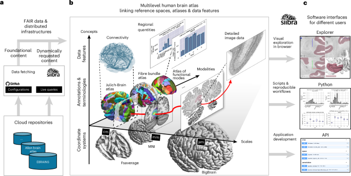
In the following, we demonstrate how key functionalities of siibra enable the implementation of a Multilevel Human Brain Atlas, linking reference atlases and comprehensive datasets from cloud resources and making the underlying data accessible and actionable for neuroscience applications (Fig. 1). All data and functionality are openly accessible. References to corresponding executable codes and online resources are provided throughout the text to ensure reproducibility.
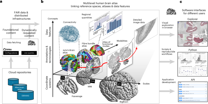
a, Contents of the multilevel atlas are curated and stored in cloud repositories, with EBRAINS (https://ebrains.eu) chosen as the main platform following the openMINDS metadata framework (https://github.com/openMetadataInitiative/openMINDS). We distinguish foundational content, predefined as versioned collections of configuration files, from dynamic content obtained at runtime using ‘live queries’ to APIs of selected cloud repositories. b, The tool suite links comprehensive data resources with reference atlases into the Multilevel Human Brain Atlas, covering different reference coordinate systems and atlas annotations which capture inter-subject variability as well as microscopic details of individual brains. Brain areas and spatial locations are linked with multimodal datasets, including microscopic images, connectivity matrices and regional quantities such as neurotransmitter receptor densities. The red arrow illustrates a typical access path from a brain area to a detailed measurement in a relevant dataset. c, The tool suite is a modular environment consisting of cloud services and installable software components. It provides access to all contents of the Multilevel Human Brain Atlas in harmonized, actionable data formats through interactive and programmatic user interfaces.
The work relies on several key concepts, whose distinctions are crucial for understanding the presented results and methodology. First, we distinguish individual reference atlases from the overarching concept of the Multilevel Human Brain Atlas. The latter combines complementary reference atlases reflecting different principles of brain organization (Fig. 1b), such as the Julich-Brain atlas (cytoarchitecture6), the deep white matter bundle atlas (fiber architecture7) or the atlas of dictionaries of functional modes (functional architecture8). Furthermore, following the AtOM ontology model9, we assume that each individual reference atlas provides a terminology of brain area definitions, often in hierarchical form. Brain areas are delineated in images or surfaces of different reference coordinate systems, resulting in annotation sets. Annotations may be given in the form of labeled or statistical maps, reflecting discrete or continuous annotations respectively. Discrete annotation sets covering the whole brain are often further denoted as parcellation maps.
The siibra tool suite is a modular environment consisting of cloud services and installable software components (Fig. 1). It includes an interactive 3D web viewer (siibra-explorer), an HTTP API (siibra-api), and a Python library (siibra-python). In either form, it assumes minimal technical requirements: The online viewer can be accessed with common web browsers, including mobile devices, and co-display user-supplied NIfTI files with atlas content without uploading them. The Python client can be installed with established package managers from https://pypi.org/project/siibra and provides a broad range of documented code examples at https://siibra-python.readthedocs.io/en/v1/examples.html.
The tool suite does not store any actual data itself. Instead, all atlas elements and data features (referred to as siibra content) are tagged with comprehensive metadata and point to published data resources (Fig. 1a). This includes reference atlases and coordinate systems, as specified in Extended Data Table 1 and Extended Data Fig. 1 for the Multilevel Human Brain Atlas, as well as a selection of multimodal datasets linked to these. Because content is separated from software, installation of the Python client requires only minimal storage space. Following a lazy data-fetching strategy, siibra downloads only requested data elements to gradually build up a configurable local data cache according to user needs. For operating the system in commercial or clinical environments with restricted network policies, the complete tool suite can be set up for offline use by cloning the configuration to a local computer, pre-populating the cache explicitly and deploying the API and viewer components as local network services.
siibra enables specifying and processing various forms of spatial locations in the brain, such as centers of gravity of measured neuroimaging signals, coordinates of functional activations and bounding boxes of regions of interest in brain tissue. These locations can be transformed between reference coordinate systems using precomputed diffeomorphic transformations, with an expected accuracy depending on the degree of nonlinear deformation in the respective region (Extended Data Fig. 2).
Locations can be assigned to brain areas defined in the reference atlases through multilayered semantic and spatial relationships, and used to retrieve multimodal data features connected to these structures. Content elements are then fetched and exposed by siibra in interoperable formats (Extended Data Fig. 3).
Due to the distributed nature of the underlying data resources, response times for data retrieval can vary depending on the location of the user and the server hosting the data. At present, most of siibra’s contents are hosted on EBRAINS, which uses storage systems on European supercomputing centers. We analyzed response times when running a typical workflow repeatedly from different geolocations, and found that the performance of siibra is usually fluid and reliable inside Europe, but could be unsatisfactory from other continents including North America, Asia and Australia (Extended Data Fig. 4).
Further details of the software architecture design are described in the Methods section.
We have developed siibra to implement a multilevel atlas of the human brain that is integrated with the EBRAINS research infrastructure (https://ebrains.eu) and provides interfaces to additional data repositories. This atlas combines reference atlases and coordinate spaces at different scales (Extended Data Table 1 and Extended Data Fig. 1) with multimodal datasets (Extended Data Table 2), supporting voxel and surface-based workflows for the whole brain while offering access to underlying data at microscopic detail.
The reference spaces include at the macroscopic scale the International Consortium for Brain Mapping (ICBM) 2009c nonlinear asymmetric multisubject and the Colin27 single-subject average templates10 as well as surface subspaces (fsaverage and fsaverage6) from the Freesurfer software suite11,12. These are combined with the BigBrain space at microstructural level13, which represents a 3D reconstruction of 7,404 histological sections from an individual postmortem human brain with an isotropic voxel resolution of 20 × 20 × 20 μm.
A central element of the Multilevel Human Brain Atlas is the Julich-Brain cytoarchitectonic atlas6, which includes probabilistic maps of more than two hundred cortical and subcortical areas for each hemisphere to date. The Julich-Brain atlas was chosen due to its rigorous multiscale approach: (1) it captures regional microstructural variance based on studies of ten postmortem brains; (2) it provides reproducible, observer-independent mapping; (3) the quality of the mapping has been validated through numerous peer-reviewed publications for all areas; and (4) it includes a microscopic template space, the BigBrain, enabling to bridge the macro scale (as represented in the MNI template) with the micro scale (as represented in BigBrain). An increasing number of mapped areas are annotated as detailed 3D maps across the entire series of BigBrain histological sections with 20-μm isotropic resolution14, supplemented by a deep learning workflow. Deep learning was instrumental in speeding up mapping in intermediate sections to achieve dense segmentation of areas over their full extent. In line with our vision of a ‘living atlas’, updates with new areas are being released at a rate of 1–2 per year. Such thorough mapping remains very time-consuming, with an estimated effort of one area per year and scientist.
The cerebral cortex in the BigBrain is further segmented by maps of six isocortical layers based on an automatic classification of 3D cortical intensity profiles15. The cytoarchitectonic maps are aligned with maps of superficial as well as long white matter bundles7,16 obtained by clustering streamlines from diffusion-weighted imaging of a large number of subjects, as well as maps of functional modules at different granularities computed from millions of fMRI scans to provide an efficient reduction and representation of BOLD signals8.
The data resources currently connected to the multilevel atlas include detailed measures of cellular (for example cell densities and staining profiles) and molecular (for example receptor densities and gene expressions) architecture, parcellation-based structural and functional connectivity from neuroimaging cohorts, and an extensive collection of high-resolution histological measurements (Extended Data Table 2).
The interactive atlas viewer siibra-explorer allows visually guided exploration of brain organization from the macroscopic scale of the brain down to individual cell bodies, and can be accessed with any web browser, including mobile touchscreen devices. It integrates very large and detailed image resources (Extended Data Table 2) into a 3D visualization of the whole brain. In addition, it embeds cross-sectional volumetric and surface views into an interactive workflow that allows us to navigate reference atlases in different coordinate systems, and to access multimodal datasets anchored to brain areas or coordinate-based locations. Cross-sectional views can be adjusted in real-time to display arbitrary oblique cutting planes and zoom levels, which is crucial, for example, for studying the cortical laminar structure of a highly folded cortical surface. Individual planar views can be maximized to obtain a large field of view, and support layering to co-display brain region maps with underlying image data.
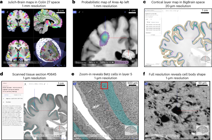
The first example concerns Betz cells in layer V of the primary motor cortex (area 4p6). The workflow starts by selecting the MNI Colin27 template and Julich-Brain as reference atlas, displayed as a cross-planar view combined with a rotatable 3D surface visualization in the corresponding reference coordinate system (Fig. 2a). By maximizing one of the cutting planes, a more focused view is obtained and can be adjusted to display an oblique sectioning angle (Fig. 2b).
a,b, Selection of area 4p in the left hemisphere from the Julich-Brain cytoarchitectonic maps6 in MNI Colin 27 space10 brings up its probability map. c, When switching to the microscopic BigBrain model as a reference space13, the level of detail is substantially increased while siibra-explorer preserves zoom and location. The parcellation is changed to cortical layer maps15, and anchored image data are revealed in the side panel. d, A 1-μm scan of a coronal whole brain section28 is selected and superimposed with the 3D model. e,f, Zooming to full resolution reveals presence and shape of individual cell bodies of Betz giant cells, clearly located in layer V. All steps can be reproduced using the following URLs: a, atlases.ebrains.eu/viewer/go/siibra_paper_2A; b, atlases.ebrains.eu/viewer/go/siibra_paper_2B; c, atlases.ebrains.eu/viewer/go/siibra_paper_2C: d, atlases.ebrains.eu/viewer/go/siibra_paper_2D; e, atlases.ebrains.eu/viewer/go/siibra_paper_2E; f, atlases.ebrains.eu/viewer/go/siibra_paper_2F.
When switching to the BigBrain template, precomputed nonlinear spatial transformations are used to preserve location and zoom level while changing the reference coordinate system (Fig. 2c). Besides inevitable trade-offs required in nonlinear cross-subject registration, as described for example in ref. 17, the precision of this coordinate translation depends on numerical limitations and the degree of nonlinear deformation between the templates in the zoomed-in region of interest (Extended Data Fig. 2).
If a particular brain area is selected in the source reference space (for example Fig. 2b), siibra-explorer tries to preserve the selection in the target space. However, a corresponding map of the region might not be available in the target space. In this case, the area selection is discarded with a corresponding notification, and the corresponding region of interest is displayed as uncharted in the target reference space.
This mechanism can be used to identify macroscopic structures in MNI space as a starting point for exploring detailed maps of cytoarchitectonic regions and cortical layers in the BigBrain15, and finding relevant microscopic image data at cellular resolution anchored to this coordinate space (Fig. 2d). The viewer allows us to select microscopic images and superimpose them with the underlying template, so that they can be explored at full spatial resolution while keeping linked with the whole brain context (Fig. 2e,f). The multilayered views can be reused by bookmarking the browser URL or downloading the underlying zoomed-in data, using the download button on the top right.
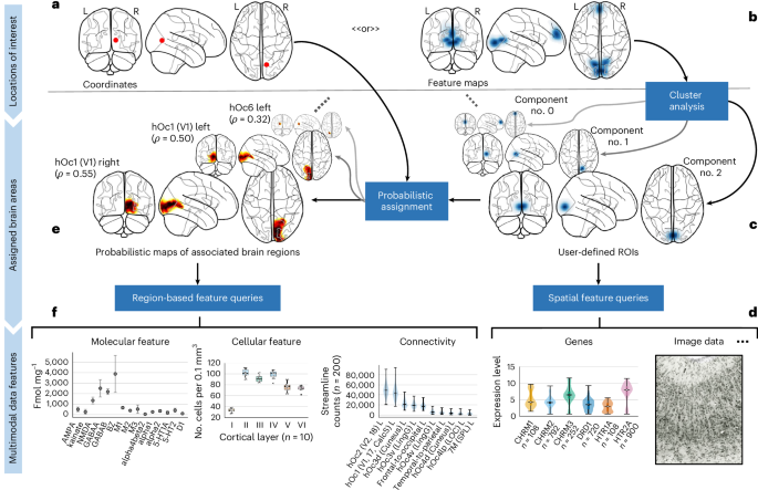
The siibra tool suite enables the assignment of user-defined regions of interest to brain areas from reference atlases and facilitates the retrieval of relevant multimodal measurements. This can be used to further characterize these regions in terms of structure, function and connectivity. In the simplest case, locations are specified as 3D coordinates in a supported reference space (Fig. 3a) using siibra’s interactive or programmatic location definition options. Associated brain regions are then assigned to brain areas from a reference atlas (Fig. 3e) utilizing the available 3D reference maps (for example functional, cyto- or fiber architectonic maps) and distinguishing between incidence, correlation and overlap of structures. In the case of statistical maps, where each brain area is typically represented by a separate image volume to account for the overlap in the maps, this might imply to retrieve, load and process several hundred image volumes. To reduce resulting download times and memory requirements, siibra uses a sparse data representation for statistical maps which minimizes the memory footprint. Associated brain areas can then be used to run data queries and find multimodal data features connected to these structures (Fig. 3f). During this process, siibra automatically resolves spatial relationships between different reference spaces and, if necessary, warps coordinates using precomputed diffeomorphic transformations.
a,b, siibra accepts user-defined location specifications in the form of reference space coordinates with optional certainty quantifiers (a) or feature maps from imaging experiments (b). c, Feature maps are typically separated into cluster components. d, Any region of interest (ROI) in image form can be used to run spatial feature queries and extract colocalized multimodal data features. e, In addition, locations of interest are assigned to brain areas using probabilistic maps from functional, cyto- or fiber architectonic reference atlases, distinguishing incidence, correlation and overlap of structures. f, Resulting associated brain areas reveal additional relevant data features. Fig. 4 provides details regarding the statistical plots in d,f. The figure can be reproduced using the tutorial notebook available in the online documentation56.
The current set of data features in the Multilevel Human Brain Atlas are grouped into the categories cellular, molecular, fibers, functional, connectivity and macrostructure (Extended Data Table 2). Their content includes combinations of image, tabular and numerical array data. Tabular data structures include cortical density measures from histological labeling, ‘fingerprints’ encoding means and s.d. of densities from tissue samples, structural and functional connectivity matrices, or external data such as gene expression levels from the Allen Human Brain microarray data18,19. Connectivity matrices are parcellation-grouped numbers and lengths of streamlines obtained from tractography of diffusion MRI data as well as correlations of resting-state and task-specific functional time series extracted from different neuroimaging cohorts. Image features include tissue sections, volumes of interest from high-resolution imaging experiments, and in vivo neuroimaging scans. Histological image data are typically anchored to the BigBrain template, as it provides the necessary level of detail to embed microscopy data.